A nucleosome (PDB code 1AOI, structure generated with PyMol) is wrapped in about 150 bases of DNA.
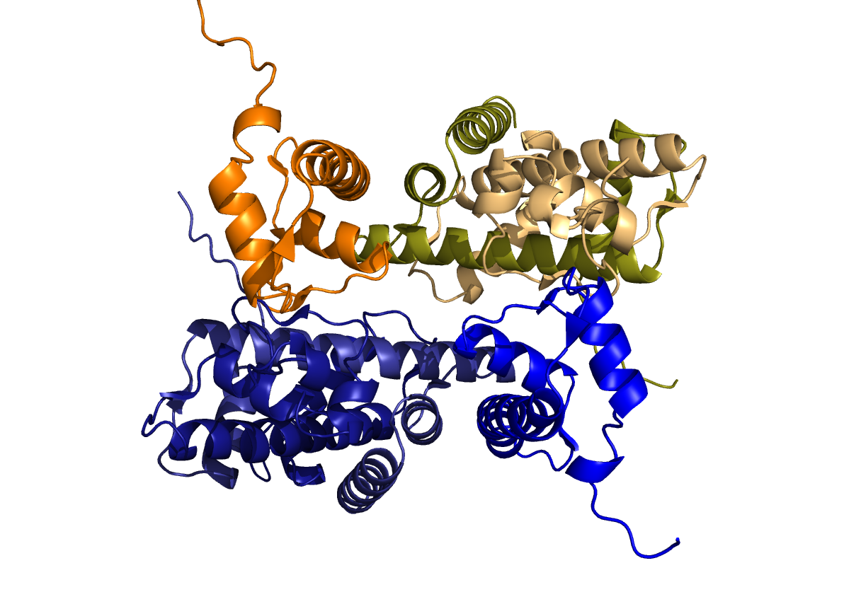
The core particle of a nucleosome is a histone octamer. It has two copies of each of the histones H2A, H2B, H3, and H4. In this image, only two subunits are shown. Despite their different appearances, the two chains have identical sequences. One simply has an alpha-helix unrolled.
Here are the other six subunits. The complex of three histones on top and the complex underneath have the same sequences and similar structures. The axis of symmetry of the histone complex runs horizontally through the middle of this image.
This image is a combination of the previous two, showing the symmetry of all eight subunits. It also shows that DNA wraps in their plane of symmetry, and that three manganese atoms that crystallized with histone are all on one side.
This model (PDB code 3C9K, made using JMol) is a possible aggregate of nucleosomes within a chromatin fiber. It is a four-helix, and might not be the real structure.


This is very cool--I need to learn how to make videos like this.
ReplyDeleteCool pictures and the video too! I like how you use the pictures from histone interactions with other things too. However, I just wish that you had more descriptions about the histone itself and where it is used. I think you are missing two blog posts too right?
ReplyDelete